2016-08-31_0001.jpg

Author: Dr Tim Bracey

2016-08-31_0001.jpg

Author: Dr Tim Bracey

2016-08-31_0001.jpg

Author: Dr Tim Bracey

2016-08-31_0001.jpg

Author: Dr Tim Bracey

2016-08-31_0001.jpg

Author: Dr Tim Bracey

2016-08-31_0001.jpg

Author: Dr Tim Bracey

22/08/2016         1:00 PM         Suffix - Tissue Types                    HI022311W/16
                                                   - Not Fully Authorised -
     1 - Gastric Biopsy                            
 
 Report sequence 1              Slides Issued                                    31/08/2016 12:10
                                          By   Dr T Bracey                               CELLPATH (Tim's laptop)
NATURE OF SPECIMEN:    2WW
Gastric polyp biopsy x 4.
CLINICAL DETAILS:
Antral polyp.
GROSS DESCRIPTION:
Four biopsies  measuring: largest 4mm, smallest 3mm. 
s4 1 NSW   PJ/ID
HISTOLOGY:
Medical Codes
** T-57000  - Stomach, NOS
             PC-TIM   - Case reported after consultation with Dr Bracey
 
Pathologists: 
Dr MEF Smith
 

14/06/2016         2:41 PM         Suffix - Tissue Types                    HI015443D/16
     1 - Duodenal Biopsy                       2 - Gastric Biopsy
     3 - Gastric Biopsy                            
 
 Report sequence 1              Authorised                                       10/08/2016 07:38
                                          By   Steve Blunden                          CELLPATH (Steve Blunden) PC
NATURE OF SPECIMEN:
1.  D2 biopsy x 4.
2.  Pylorus biopsy x 2.
3.  Gastric polyp biopsy x 1.
CLINICAL DETAILS:
IDA.  Weight loss.  CLO test.
GROSS DESCRIPTION:
1.  Four biopsies, the largest measuring 4mm and the smallest 2mm.  S4 1B NS AM
2.  Two biopsies, the larger measuring 3mm and the smaller 2mm.  S2 1B NS AM
3.  One biopsy, measuring 2mm.  S1 1B NS AM
HISTOLOGY:

(1) This is small bowel mucosa.  There is a regular crypt and villous architecture with an intact brush border.  There is no evidence of inflammation, coeliac disease, parasitic infection or dysplasia.


(2 & 3) This is antral-type gastric mucosa showing an irregular architecture with marked foveolar hyperplasia with infolding of the mucosa.  There is mild active chronic inflammation and granulation tissue containing bizarre fibroblasts is seen.  Intestinal metaplasia, Helicobacter or atrophy are not seen.  There are regenerative changes but no evidence of dysplasia.


The appearances for both bopsies are those of a regenerative polyps. In specimen 2 (pylorus) there is some nuclear atypia which is most probably regenerative in nature.  However interpretation can be difficult in the context of inflammation and therefore early re-biopsy is recommended.


Diagnostic Summary

(1) Duodenal biopsies - Normal.


(2 & 3) Gastric polyp - regenerative polyp.  No evidence of Helicobacter or dysplasia but early re-biopsy recommended (see main report).
Reported by: Professor Mohammad Ilyas


Stomach

Polyps

Hyperplastic polyp


Reviewers: Elliot Weisenberg, M.D. (see Reviewers page) 
Revised: 8 August 2012, last major update August 2012
Copyright: (c) 2003-2012, PathologyOutlines.com, Inc.

General
=========================================================================

● Also called inflammatory, regenerative polyps
● Different from colonic hyperplastic polyps but similar to colonic inflammatory pseudopolyps
● Non-neoplastic, represents about 75% of all gastric polyps
● Associated with background mucosal disease in 85% of cases, particularly chronic gastritis with glandular atrophy and intestinal metaplasia; H. pylori gastritis, chemical (reactive) gastropathy, thus, endoscopists should also biopsy surrounding mucosa to evaluate underlying gastric abnormalities
● Also associated with hypochlorhydria and hypergastrinemia
● Multiple polyps associated with autoimmune gastritis
● Usually age 50+
● Rare/no malignant potential by itself, although present in 20% of stomachs resected for carcinoma (chronically inflamed and atrophic mucosa tends to form hyperplastic polyps and to degenerate into malignancy)
● Hyperplastic polyps with even low grade dysplasia may have significant risk for associated carcinoma (Hum Pathol 2002;33:1016)
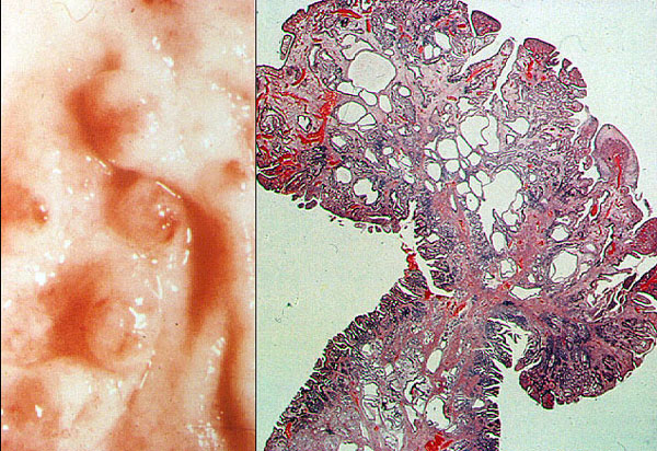
Gross description
=========================================================================

● Small (mean 1.4 cm, range 0.5 to 2 cm), sessile, multiple (20%), 60% in antrum
● Smooth or slightly lobulated
● Central umbilication common, more proximal when associated with autoimmune gastritis 
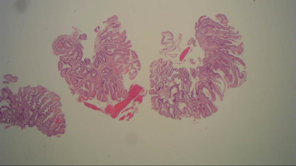
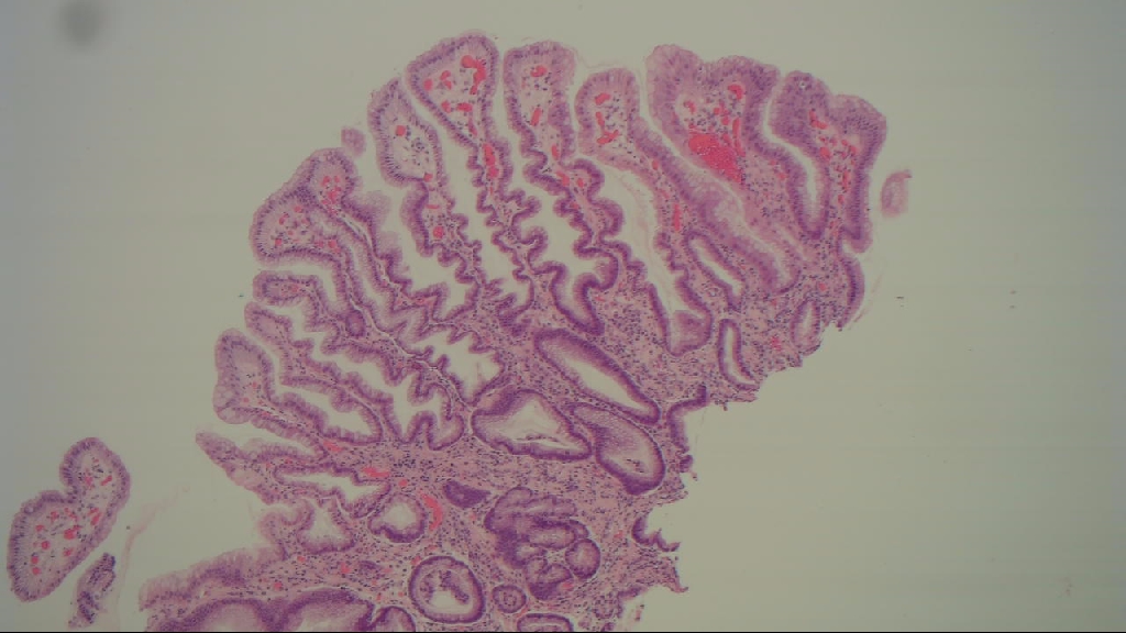
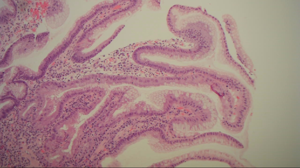
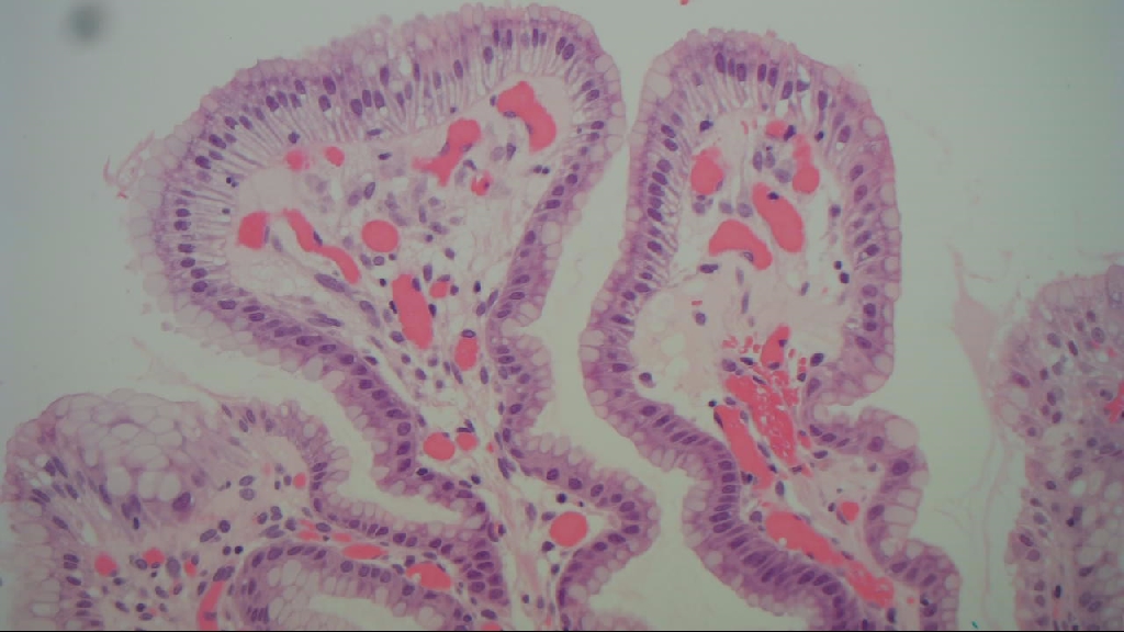
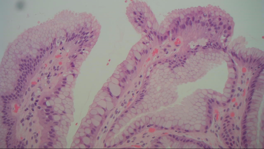
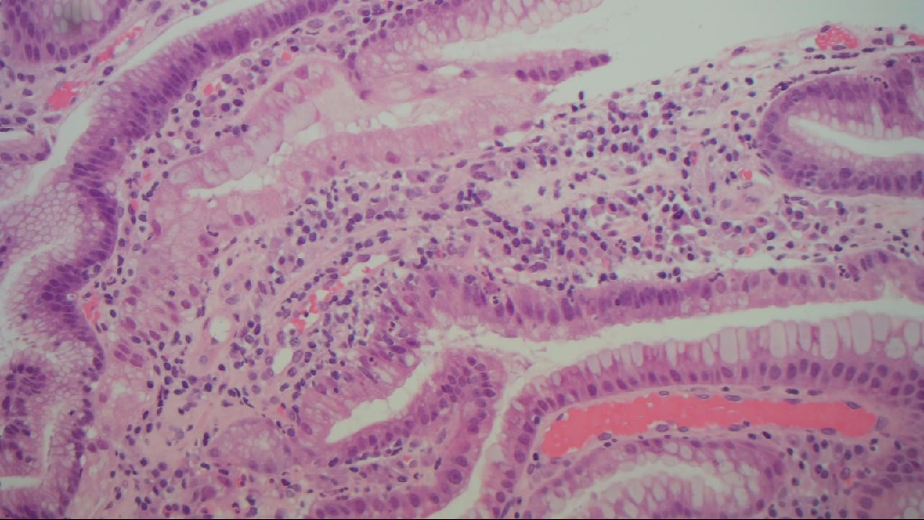
Micro description
=========================================================================

● Elongated, tortuous or dilated gastric foveola with pyloric or fundic glands (Am J Surg Pathol 2001;25:500)
● Lamina propria has inflammatory cells, scattered smooth muscle bundles, edema, patchy necrosis
● Associated with chronic, active (H. pylori), reactive gastritis, and atrophic autoimmune gastritis
● Rarely foamy macrophages
● Surface mucosa may be regenerative, but dysplasia in only 4%
● Focal intestinal metaplasia in 16%
● May regress when H. pylori gastritis is treated

Micro images
=========================================================================


Hyperplastic polyps